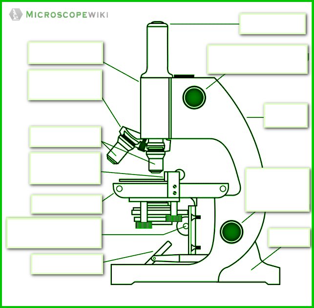
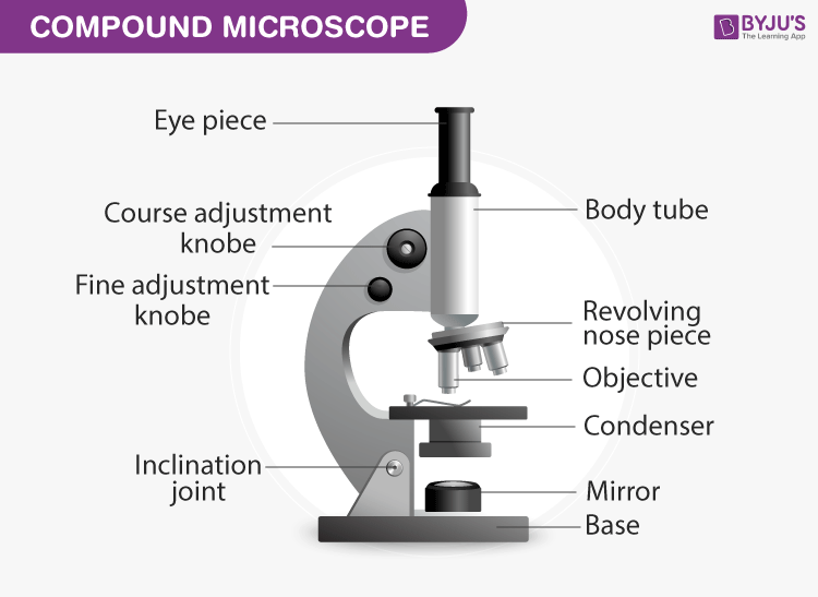
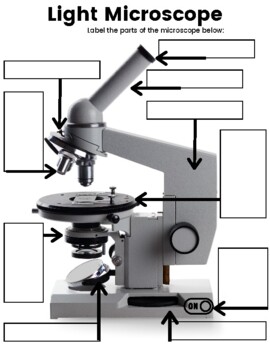
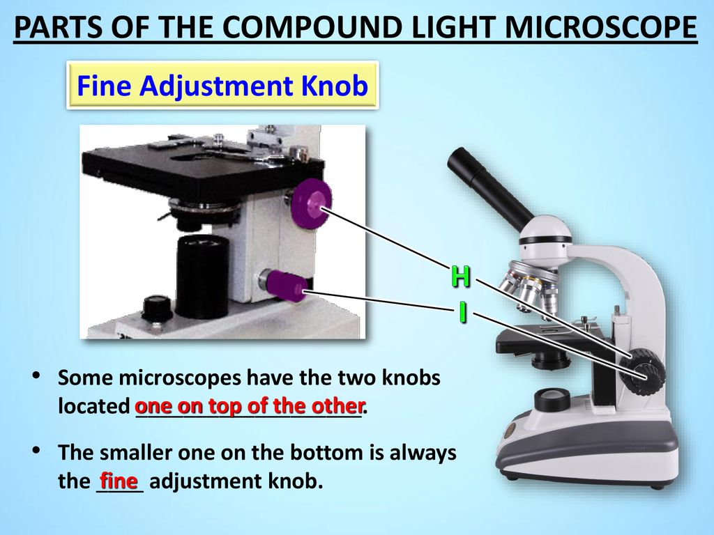
41 microscope diagram worksheet
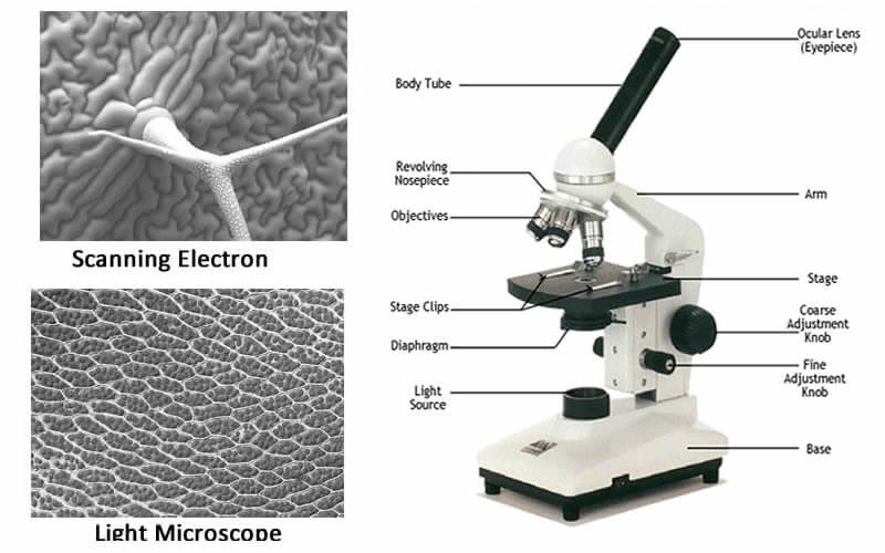
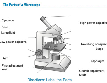
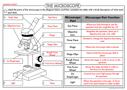
PDF Parts of the Microscope Study Sheet - 0.tqn.com 6.Objectives The lens or system of lenses in a microscope that is nearest the object being viewed 7.Diaphram Located on the stage, adjusts the amount of light passing into the slide 8.Base The bottom of the microscope, used for support 9.Arm Supports upper part of the microscope 10.Stage Small platform where the specimen is mounted for examination rsscience.com › stereo-microscopeParts of Stereo Microscope (Dissecting microscope) – labeled ... Labeled part diagram of a stereo microscope Major structural parts of a stereo microscope. There are three major structural parts of a stereo microscope. The viewing Head includes the upper part of the microscope, which houses the most critical optical components, including the eyepiece, objective lens, and light source of the microscope.
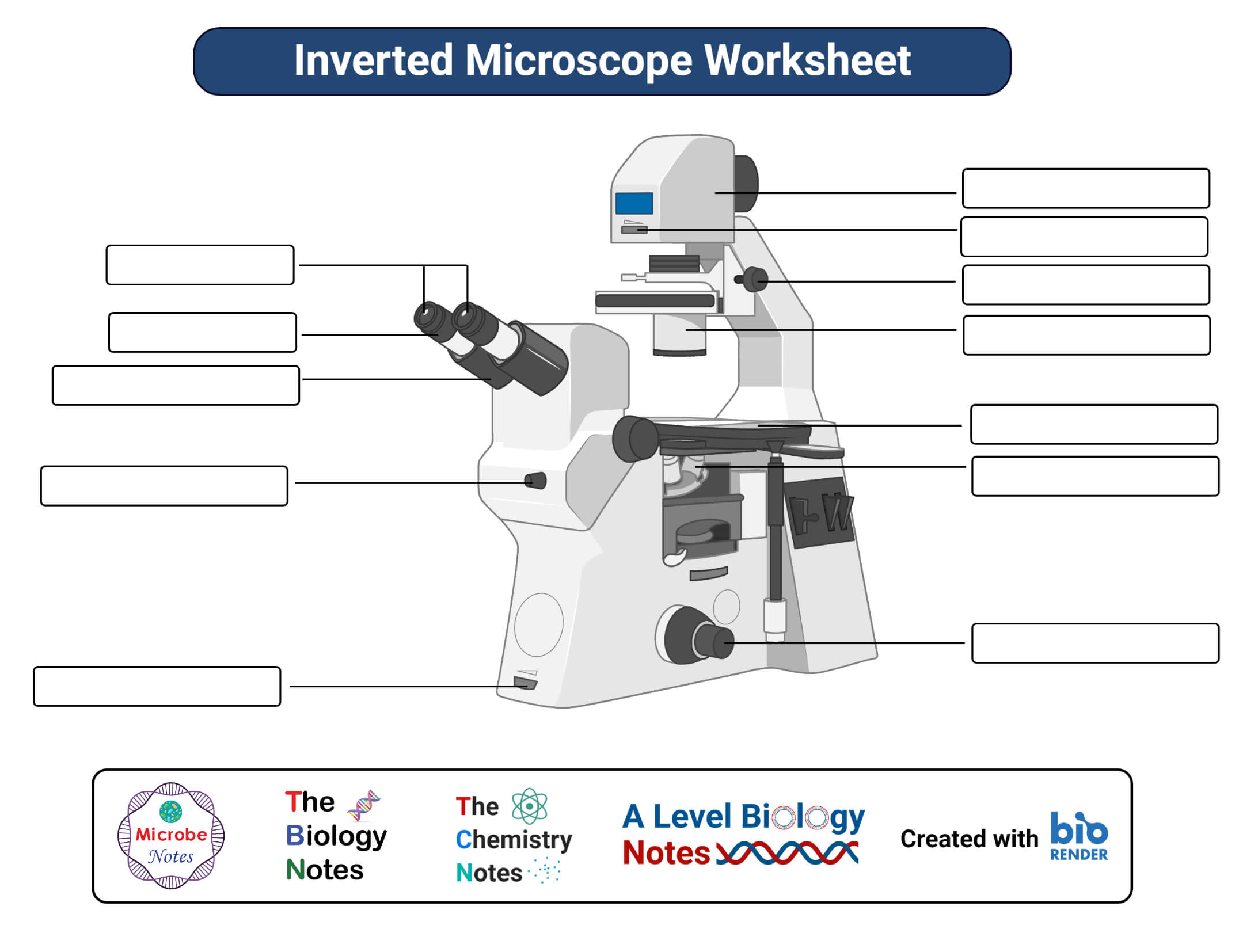
Inverted Microscope- Definition, Principle, Parts, Labeled Diagram ... The working principle of the inverted microscope is basically the same as that of an upright light microscope. They use light rays to focus on a specimen, to form an image that can be viewed by the objective lenses. However, in the inverted microscope, the light source and the condenser are found on top of the stage pointing down to the stage.

Microscope diagram worksheet
lzsgec.crabsfamily.shop › blood-under-microscopeBlood under microscope - lzsgec.crabsfamily.shop This worksheet can be used with the Virtual Microscope where students can place specimens on a stage and use coarse and fine adjustment knobs to magnify up to 100x.. Generally, I have my students practice with real microscopes , starting with a basic tutorial lab where they focus on the letter “e.” Microscope Lab Worksheet Name I.... - Course Hero Go find information about both types of microscopy on the Internet, then briefly compare the advantages and disadvantages of each type of imagery for studying skin cells and their organelle structures. Please reference the two images provided on the lab page in your answer. 1 Microscope Lab Worksheet PDF Microscope Calculations - Vancouver Community College When looking into a microscope, you will see a lit circular area. The distance across the center of the circle is referred to as the diameter of field of view (dFOV). To calculate the dFOV, you will need to place a transparent ruler on the microscope stage and measure the dFOV under low power. Looking at the diagram below, we can see that
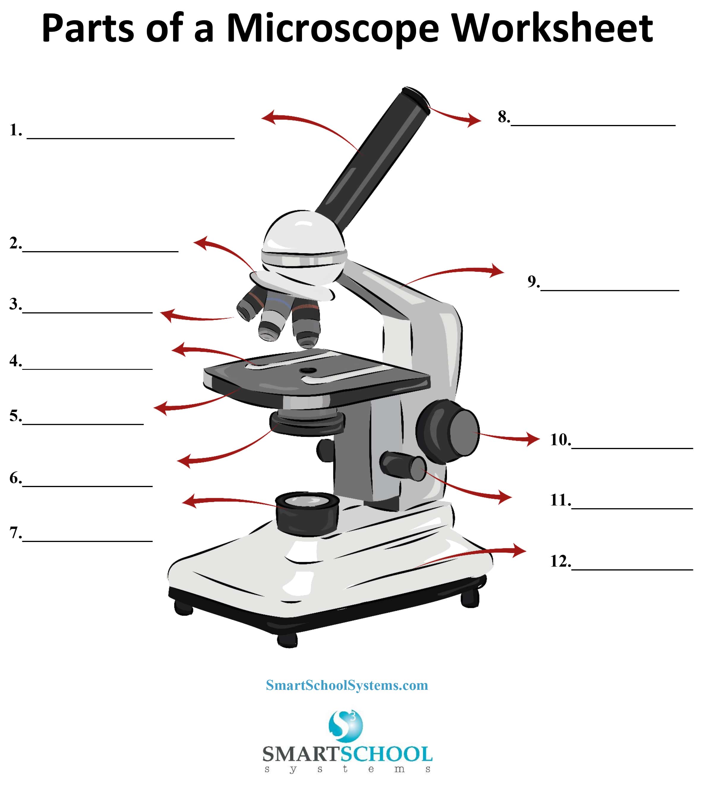
Microscope diagram worksheet. PDF Microscope Worksheet Microscope Worksheet I. How Can a Microscope Help Us Study Living Things? Microscope: an instrument that makes things look bigger Biology: the study of living things. Some living things are so small we can't see them with the naked eye alone. Many of these are one celled organisms called PROTOZOA [pro tuh ZO uh] and BACTERIA. PDF Name: Class: Microscope Worksheet - MRS. BOGUSLAW'S WEBPAGE Stage- a platform on top of the base of the microscope on which specimen are placed Stage Clip- clips on top of stage that allow you to secure the speciman or slide. Revolving Nosepiece- a portion of the microscope that allows you to switch back and forth between lens powers. Power Switch- on/off button that powers the light. Microscope Parts Diagram - Printable About this Worksheet This is a free printable worksheet in PDF format and holds a printable version of the quiz Microscope Parts Diagram. By printing out this quiz and taking it with pen and paper creates for a good variation to only playing it online. microbenotes.com › plant-cellPlant Cell- Definition, Structure, Parts, Functions, Labeled ... Feb 16, 2022 · Figure: Labeled diagram of plant cell, created with biorender.com. The typical characteristics that define the plant cell include cellulose, hemicellulose and pectin, plastids which play a major role in photosynthesis and storage of starch, large vacuoles responsible for regulating the cell turgor pressure.
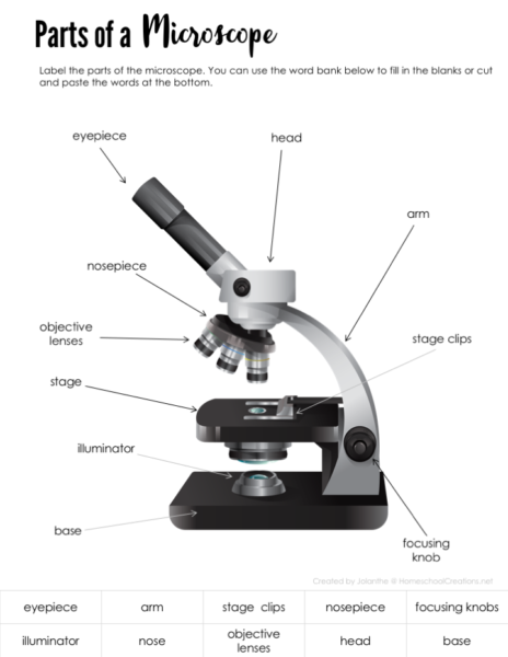
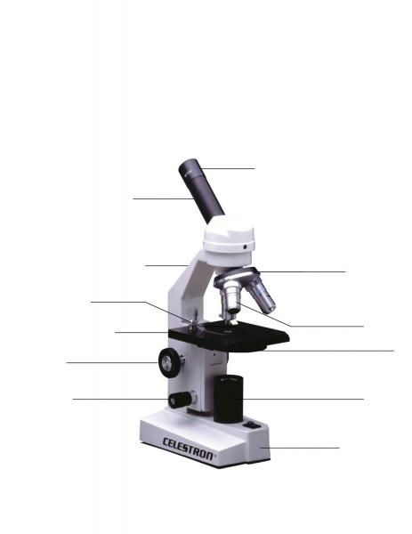
Science 10 Microscope Worksheet: - St. Clair County School District If you draw a diagram which is 36mm, what is the magnification? An object has an actual length of 0.025mm. If you use a scale of 1:1000, what will be the size of the drawing? An organism has an actual length of 0.033mm. If you use a scale of 1:250, what will be the size of the drawing? We see very small things by using a microscope to look at them. Labeling the Parts of the Microscope | Microscope World Resources Labeling the Parts of the Microscope This activity has been designed for use in homes and schools. Each microscope layout (both blank and the version with answers) are available as PDF downloads. You can view a more in-depth review of each part of the microscope here. Download the Label the Parts of the Microscope PDF printable version here. Microscope Diagram Worksheets - K12 Workbook Displaying all worksheets related to - Microscope Diagram. Worksheets are The microscope parts and use, Parts of the light microscope, Powerpoint work the microscope diagram, Label parts of the microscope answers, Parts of the microscope quiz, Parts of the microscope study, An introduction to the compound microscope, Plant and animal cells. PDF Parts of a Microscope Printables - Homeschool Creations The lenses in a microscope make items appear smaller. How many parts of a microscope can you identify? Can you show the arm, stage, eyepiece, head, objective lens, illuminator, nosepiece, and stage clips? Where is the safest place to hold or carry a microscope? Which part of the microscope holds the specimen slide in place? Why do we use ...
Simple Microscope - Diagram (Parts labelled), Principle, Formula and Uses Simple Microscope Diagram with Labels Simple Microscope - Student Free Worksheet Frequently Asked Questions Q 1. How many lenses does a simple microscope have? Simple microscope has one objective lens for magnifying the specimen Q 2. When was the simple microscope invented? The simple microscope was invented by Anton van Leeuwenhoek in 1675 Q 3. FREE Microscope Parts Worksheet - This free microscope diagram ... Print a microscope diagram, microscope worksheet, or practice microscope quiz in order to learn all the parts of a microscope. Yara Qerem. Biology. Teaching Cells. Science Ideas. Upper Elementary Resources. 5 easy steps for focusing a compound light microscope. Additional tips and tricks for purchasing microscopes included. Compound Light Microscope Diagram Worksheet - Google Groups In this microscopes worksheet students compare and contrast the characteristics and uses of pale light microscope and electron microscope. Always return to match up through a diagram. Remove your f... PDF The Microscope Parts and Use - Plainview the year 1590. The compound microscope uses lenses and light to enlarge the image and is also called an optical or light microscope (vs./ an electron microscope) . The simplest optical microscope is the magnifying glass and is good to about ten times (10X) magnification. The
microscope diagram worksheet Microscope parts compound worksheet diagram printable worksheets quiz grade biology science labeling answers sketch labeled blank tests label light learning. Microscope parts science worksheets word cells worksheet middle activity vocabulary tools quiz blank biology printables searches mania grade answers blood. Cell animal plant cells electron ...
Microscope Labeling - The Biology Corner Microscope Labeling Microscope Labeling Microscope Use: 15. When focusing a specimen, you should always start with the _____________ objective. 16. When using the high power objective, only the _______________ knob should be used. 17. The type of microscope used in most science classes is the ______________ microscope. 18.
PDF Microscope calculations - MissHaynes-Science Microscope calculations 1. Converting between units You only need to remember two sums to convert between any units. Basic conversions Complete the table below Example 1 Example 2 Convert 2350nm Convert 143mm 2350nm ÷ 1000 = 0.2350μm 143mm x 1000 = 143,000μm Millimetre Micrometre Nanometre 1580 0.23 174 3
Microscope Labeling - The Biology Corner 1) Start with scanning (the shortest objective) and only use the COARSE knob . Once it is focused… 2) Switch to low power (medium) and only use the COARSE knob . You may need to recenter your slide. Once it is focused.. 3) Switch to high power (long objective).
PDF AN INTRODUCTION TO THE COMPOUND MICROSCOPE - Rowan University On your worksheet below calculate the total magnification for each ocular/objective combination on your microscope. List the magnification for each objective on your microscope below. EYEPIECE/ OCULAR X OBJECTIVE = TOTAL MAGNIFICATION Stage: The surface or platform on which you place the microscope slide is the stage.
Microscope Diagram Worksheet - Stock Walker Microscope Diagram Worksheet - The Microscope Create A Labelled Diagram Teaching Resources :. Microscope g stage d nosepiece j base e. When you can identify a part of the microscope place the . Write the letter on the line that represents each part of the microscope. Use the following terms to correctly label the microscope: Go to a microscope ...
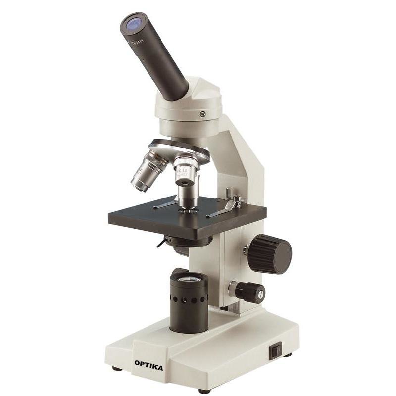
microbenotes.com › parts-of-a-microscopeParts of a microscope with functions and labeled diagram Apr 19, 2022 · Figure: Diagram of parts of a microscope. There are three structural parts of the microscope i.e. head, base, and arm. Head – This is also known as the body. It carries the optical parts in the upper part of the microscope. Base – It acts as microscopes support. It also carries microscopic illuminators.
rockyourhomeschool.net › microscope-worksheetsFree Microscope Worksheets for Simple Science Fun for Your ... Parts of a Microscope . The first worksheet labels the different parts of a microscope, including the base, slide holder, and condenser. If you have a microscope, compare and contrast this worksheet to it. Also, your kids can color this microscope diagram in and read the words to each part of the microscope.
Learn About Microscopes With Fun, Free Printables - ThoughtCo The letters of the microscope parts are all mixed up on this worksheet. Students should use the clues to figure out the correct word or words and write them on the blank lines provided. Alphabet Activity Beverly Hernandez
Microscope Diagram Teaching Resources | Teachers Pay Teachers Parts of a Microscope Overview Reading Comprehension and Diagram Worksheet by Teaching to the Middle 4.8 (24) $1.50 PDF This passage briefly describes microscopes and their parts (900-1000 Lexile). 14 questions (matching and multiple choice) assess students' understanding. Students label a diagram of 6 parts of a microscope.
PDF Label parts of the Microscope: Answers Label parts of the Microscope: Answers Coarse Focus Fine Focus Eyepiece Arm Rack Stop Stage Clip . Created Date: 20150715115425Z ...
› langarts › partsofSubject Pronouns Worksheets - Easy Teacher Worksheets Subject pronouns are used as the subject of action words. The usually take the place of regular nouns. They really give mean to thing, person or place. They point to the meaning of the passage or reading. When the sentence is restricted to a few lines, this personal noun helps pin point the object or person that is under the microscope.
› watchThe wacky history of cell theory - Lauren Royal-Woods View full lesson: discovery isn't as simple as one good experiment. The weird and wonder...
Microscope Diagram - Free Printable Tests and Worksheets | Biology ... Microscope Diagram (Grade 8) - Free Printable Tests and Worksheets Write the letter on the line that represents each part of the microscope. Microscope G Stage D Nosepiece J Base E Help Teaching 10k followers More information FREE Printable Microscope Diagram Worksheet - great for learning the parts of a microscope or use as a quiz
PDF Parts of the Light Microscope - Science Spot B. NOSEPIECE microscope when carried Holds the HIGH- and LOW- power objective LENSES; can be rotated to change MAGNIFICATION. Power = 10 x 4 = 40 Power = 10 x 10 = 100 Power = 10 x 40 = 400 What happens as the power of magnification increases?
PDF Microscope Calculations - Vancouver Community College When looking into a microscope, you will see a lit circular area. The distance across the center of the circle is referred to as the diameter of field of view (dFOV). To calculate the dFOV, you will need to place a transparent ruler on the microscope stage and measure the dFOV under low power. Looking at the diagram below, we can see that
Microscope Lab Worksheet Name I.... - Course Hero Go find information about both types of microscopy on the Internet, then briefly compare the advantages and disadvantages of each type of imagery for studying skin cells and their organelle structures. Please reference the two images provided on the lab page in your answer. 1 Microscope Lab Worksheet
lzsgec.crabsfamily.shop › blood-under-microscopeBlood under microscope - lzsgec.crabsfamily.shop This worksheet can be used with the Virtual Microscope where students can place specimens on a stage and use coarse and fine adjustment knobs to magnify up to 100x.. Generally, I have my students practice with real microscopes , starting with a basic tutorial lab where they focus on the letter “e.”



















Post a Comment for "41 microscope diagram worksheet"